Digital X-Rays in Coral Springs, FL: A Patient Guide
Digital X-rays in Coral Springs, Florida, provide clear, detailed images that help diagnose concerns early for patients at Delight Dental Smiles Coral Springs. This modern dental technology reduces radiation exposure, speeds up appointments, and supports accurate treatment planning. Below, find answers to common questions, how the process works, and what to expect during an exam.
Digital X-Rays Explained
Digital dental X-rays use an electronic sensor to capture images of your teeth, bone, and supporting structures. Instead of developing film, the image appears instantly on a computer screen. This allows our dentists to enlarge areas, adjust contrast, and highlight details for a more precise evaluation. Compared with traditional film, digital systems often use 50% to 80% less radiation while delivering high-quality images.
Common Image Types
- Bitewing images show the upper and lower back teeth to check for cavities between teeth and bone levels.
- Periapical images focus on a single tooth from crown to root to assess infection, fractures, or bone changes.
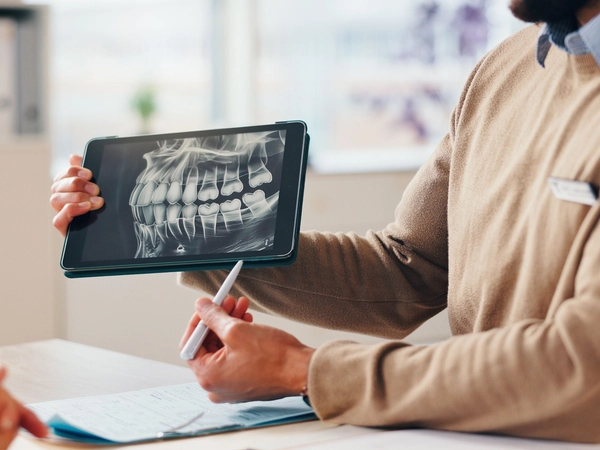
- Panoramic images provide a broad view of the jaws, sinuses, and developing teeth for orthodontic or surgical planning.
- Occlusal images show a wider area of the upper or lower arch to evaluate growth, cysts, or extra teeth.
Benefits of Digital X-Rays
- Low radiation exposure: Digital sensors reduce exposure while maintaining diagnostic quality.
- Fast results: Images appear in seconds, helping streamline your visit.
- Accurate diagnosis: Enhanced images reveal decay, gum disease, and hidden problems early.
- Better monitoring: Stored images allow easy comparison over time to track changes.
- Comfortable process: Slim sensors and fewer retakes improve the experience for many patients.
- Eco-friendly workflow: No chemical processing or film waste is needed.
How Digital X-Rays Work
- Sensor placement: A small sensor is positioned in the mouth to capture the area of interest.
- Quick exposure: The X-ray unit takes a brief exposure, often less than a second.
- Instant imaging: The picture appears on screen immediately for review and explanation.
- Analysis and planning: our dentists will evaluate findings and discuss next steps if treatment is needed.
- Secure storage: Images are saved for future comparisons and ongoing care.
What to Expect
Most appointments begin with a review of your health history and a conversation about symptoms or concerns. You will wear a protective lead apron, remove any removable metal objects, and bite gently on a sensor while images are taken. Each exposure is quick. Many patients finish a standard set of bitewing images in a few minutes.
How often digital X-rays are needed depends on your oral health, age, and risk for cavities or gum disease. New patients often receive a comprehensive set to establish a baseline. Patients with low risk may need bitewings every 12 to 24 months, while higher-risk patients may benefit from more frequent images. Children and teens may need images more often due to changing teeth and faster cavity progression. If you are pregnant, elective X-rays are usually postponed; urgent care X-rays can be done safely with shielding.
Digital X-rays help detect issues that are not visible during a visual exam, such as cavities between teeth, infections at the root tip, cracks, bone loss from periodontal disease, and problems beneath existing fillings or crowns. Early detection can reduce the complexity and cost of future treatment.